Goals
- Know myotomy technique from Exercise #3.
- Know why a partial esophageal wrap (fundoplication) is indicated for an esophagogastric
myotomy for Achalasia. - Know the technique for anterior fundoplication (Dor procedure).
Equipment
- Goat mediastinal tissue block. Fresh from local abattoir. Refrigerate. Consists of heart, lungs, esophagus, trachea, diaphragm, thoracic aorta and esophagogastric junction with attached proximal one third of the stomach.
- Maloney tapered bougie—Size 54 French for human adult. Size 45 French for goat model.
- 17-inch x 24-inch plastic cutting board with single screw placed in the center edge of each
end of longest dimension. - Lubricant-tube of K-Y jelly or Lubriderm.
- One pair of Metzenbaum scissors.
- Surgical forceps.
- 0 Ethibond sutures.
- 2-0 Ethibond sutures.
- Suture scissors.
- Curved hemostat.
- Babcock clamp.
- Needle holder.
- Spool of heavy twine.
Preparation
- Attach the mediastinal tissue block to the cutting board with heavy twine passed through the proximal esophagus and trachea. Do not obstruct the esophageal lumen. Tie this suture around the screw at the end of the cutting board. Place a second heavy twine through the distal diaphragmatic crura and tie around the screw at the distal end of the cutting board.
- See Lesson #2, Step 2, for preparation of the goat stomach to simulate the human stomach.
- Pass a lubricated esophageal bougie the full length of the esophagus.
Discussion
Lesson 5. Dor Anterior Partial Fundoplication
1. Pass an esophageal bougie by mouth through the esophagus and into the proximal stomach.
2. Describe the location and the rationale for dividing the vasa brevia between the gastric antrum and the spleen.
3. Identify the left and right crura of the diaphragm.
4. Make a 4 cm longitudinal incision through the two muscular layers of the bougie distended distal esophagus using the “belly” of a scalpel with a #10 blade. Carry this incision across the esophagogastric junction through the muscle layer of the stomach for a distance of 1.5 cm. Do not incise the submucosal/mucosal layer of the stomach.
5. Using a curved hemostat, Metzenbaum scissors and surgical forceps, elevate the esophageal muscular layers off of the submucosal layer of the esophagus for 1 cm lateral and medial to the myotomy. This will reduce the likelihood of the myotomy closing again.
6. Grasp the gastric antral tissue with a Babcock clamp and bring it anterior to the esophagogastric myotomy.
7. Place three 2-0 Ethibond sutures between the muscle layer on the patient’s left side of the myotomy and through the adjacent anterior fundic wrap and tie in place.
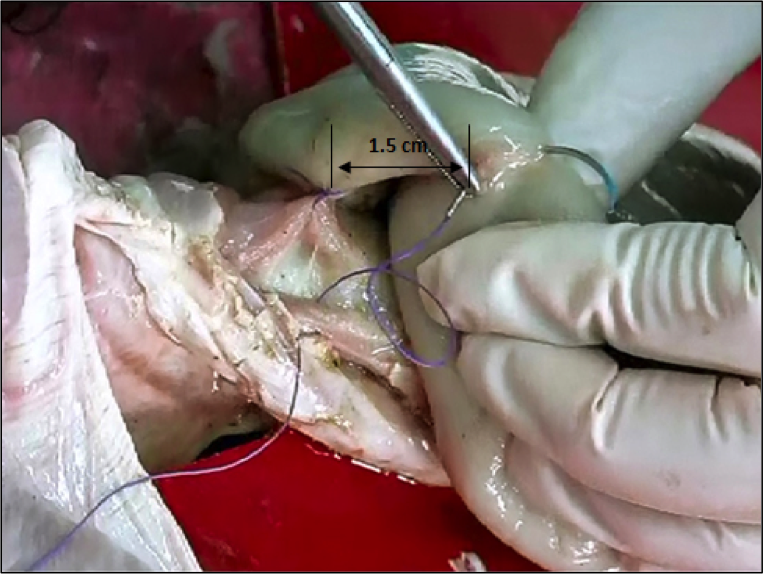
8. Place three additional 2-0 Ethibond sutures between the muscle layer on the patient’s right side of the myotomy and through the adjacent anterior fundic wrap (see below) and tie in place. Allow 1.5 cm distance between the left and right rows of suture on the anterior fundic flap. This will help hold the myotomy open.

Figure 5-1: Dor Fundoplication
9. Place two 2-0 Ethibond sutures through the right crus, the posterior wall of the stomach and the adjacent anterior fundic wrap.
10. Place two 2-0 Ethibond sutures through the left crus and the adjacent fundus. These sutures will serve to hold the partial wrap below the diaphragm.
Exercise Lesson 5.1
Exercise 5-1. Dor Partial Fundoplication 1
Videos may take a moment to load depending on your connection speed.
Video Lesson 5.2
Lesson 5-2. Dor Partial Fundoplication 2
Videos may take a moment to load depending on your connection speed.
Video Lesson 5.3
Lesson 5-3. Dor Partial Fundoplication 3
Videos may take a moment to load depending on your connection speed.