Goals
- Learn to use the “belly” of the scalpel to make a longitudinal incision in the outer muscular layers of the bougie-‐‑distended esophagus. Recognize the underlying submucosal layer and avoid incising it.
- Learn the technique of elevating the muscular layer for one third of the diameter of the esophagus at the site of the myotomy.
- Learn the technique of recognizing and repairing an inadvertent entry into the esophageal lumen during the myotomy.
Equipment
- Goat mediastinal tissue block. Fresh from local abattoir. Refrigerate. Consists of heart, lungs, esophagus, trachea, diaphragm, thoracic aorta and esophagogastric junction with attached proximal one third of the stomach.
- Maloney tapered bougies—Size 45 French.
- 17 inch x 24inch plastic cutting board with single screw placed in the center edge of each end of longest dimension.
- Lubricant—tube of K-Y jelly or Lubriderm.
- Scalpel with #10 blade.
- Curved small hemostat.
- Metzenbaum scissors.
- Surgical forceps.
- 3-0 absorbable suture.
- Needle holder.
- Heavy twine.
Preparation
- Attach mediastinal tissue block to the cutting board with heavy twine passed through the proximal esophagus and trachea. Do not obstruct the esophageal lumen. Tie this suture around the screw at the end of the cutting board. Place a second heavy twine through the distal diaphragmatic crura and tie around the screw at the distal end of the cutting board.
- See Lesson #2, Step 2, for preparation of the goat stomach to simulate the human stomach.
- Pass a lubricated esophageal bougie the full length of the esophagus.
Discussion
Esophageal Myotomy
Note: This exercise is useful to teach as it demonstrates the basic surgical maneuver of myotomy which can be repeated multiple times on the same specimen.
1. Make a 2 cm. longitudinal incision through the two muscular layers of the bougie distended esophagus using the “belly” of a scalpel with a #10 blade.
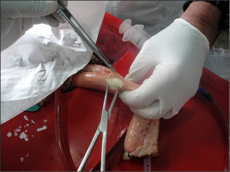
2. Using a curved hemostat, Metzenbaum scissors and surgical forceps, elevate the esophageal muscular layers off of the submucosal layer for one third of the esophageal circumference.

Figure 3.1: Esophageal Myotomy
Esophagogastric Myotomy
1. Make a 4 cm longitudinal incision through the two muscle layers of the bougie-distended distal esophagus.
2. Extend the myotomy for a distance of 1.5 centimeters onto the gastric cardia.
3. Using a curved hemostat, Metzenbaum scissors and surgical forceps, elevate the esophageal muscular layers off of the submucosal layer for one third of the esophageal circumference. Using the same instruments, elevate the gastric muscle layers adjacent to the myotomy just enough to assure a one centimeter separation of the muscle layers at the site of the gastric myotomy.
Closure of an Inadvertent Entry into the Esophageal Lumen while Doing a Myotomy
1. Recognize the site of the lumen entry, grasp the tissue edges gently with forceps and close with absorbable 3-0 suture.
Video Exercise 3.1
Exercise 3-1. Myotomy
Video Exercise 3.2
Exercise 3-2. Esophagomyotomy 1
Video Exercise 3.3
Exercise 3-3. Esophagomyotomy 2