Goals
- Be able to identify the esophagogastric junction and the left and right crura of the diaphragm.
- Understand how the total wrap of the gastric fundal tissue replicates the normal gastric anti-reflux mechanism. It holds the esophagogastric junction below the diaphragm and creates a high‐pressure zone at the esophagogastric junction.
- Understand the role of the bougie. It avoids too tight a wrap.
Equipment
- Goat mediastinal tissue block. Fresh from local abattoir. Refrigerate. Consists of heart, lungs, esophagus, trachea, diaphragm, thoracic aorta and esophagogastric junction with attached proximal one third of the stomach.
- Maloney tapered bougie- Size 54 French for human adult. Size 45 French for goat model.
- 17 inch x 24 inch plastic cutting board with single screw placed in the center edge of each
end of longest dimension. - Lubricant-tube of K-Y jelly or Lubriderm.
- One pair of Metzenbaum scissors.
- Surgical forceps.
- 0 Ethibond sutures.
- 2-0 Ethibond sutures.
- Penrose drain.
- Kelly clamp.
- Suture scissors.
- Babcock clamp.
- Needle holder.
- Suture scissors.
- Spool of heavy twine.
Preparation
- Attach the mediastinal tissue block to the cutting board with heavy twine passed through the proximal esophagus and trachea. Do not obstruct the esophageal lumen. Tie this suture around the screw at the end of the cutting board. Place a second heavy twine through the distal diaphragmatic crura and tie around the screw at the distal end of the cutting board.
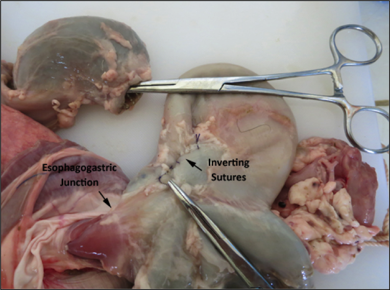
- The goat stomach has four compartments. It is useful to separate one of these compartments, the reticulum, from the rumen, omasum and abomasum in order to more closely simulate the human gastric anatomy (see next page). The Kelly clamp is attached to the thick, muscular reticulum that has been surgically removed. The incision in the rumen near the esophagogastric junction is closed with inverting sutures. The proximal portion of the rumen then becomes anatomically similar to the fundus of the human stomach.
Note: this technique of separating the reticulum from the rumen is used in Exercises #3, #4, and #5 to prepare the goat stomach model.

Figure 2.1: Preparation of the Goat Stomach to Simulate the Human Stomach
3. Pass a lubricated esophageal bougie the full length of the esophagus into the proximal stomach.
Discussion
Transabdominal Nissen Fundoplication
1. Pass an esophageal bougie by mouth through the esophagus and into the proximal stomach.
2. Describe the location and the rationale for dividing the vasa brevia between the gastric antrum and the spleen.
3. Identify the left and right crura of the diaphragm.
4. Create by blunt dissection a circumferential space around the intra-abdominal segment of the distal esophagus and then pass a Penrose drain to retract the esophagus while enlarging the space adequate to accommodate the gastric fundal wrap.
5. Grasp the gastric antral tissue and pass posterior to esophagogastric junction through the space created by Step #2.
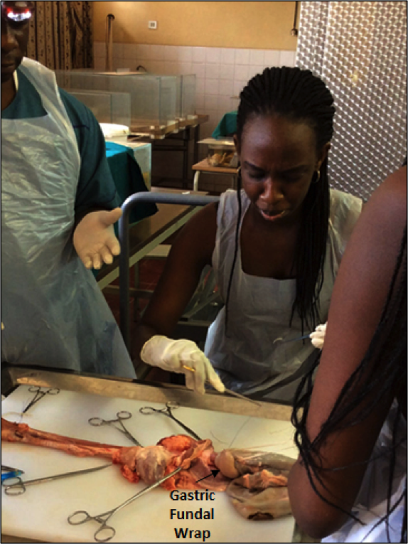
6. Bring the end of “wrap” anterior to the esophagogastric junction by grasping it with a Babcock clamp and suture to the gastric antrum on the patient’s left side with three 2-0 Ethibond sutures for a total wrap length of 1.5 cm (see below).

Figure 2.2: Nissen Fundoplication
7. Retract the bougie into the thoracic esophagus and evaluate the crural space posterior to the wrap. If further closure of the crural space is indicated, place crural sutures of 2-0 Ethibond with the bougie retracted. Intermittently advance the bougie to “size” the space between the esophagus and the diaphragmatic crura. When appropriate crural sutures have been placed, the space should be just tight enough to admit one finger with ease.
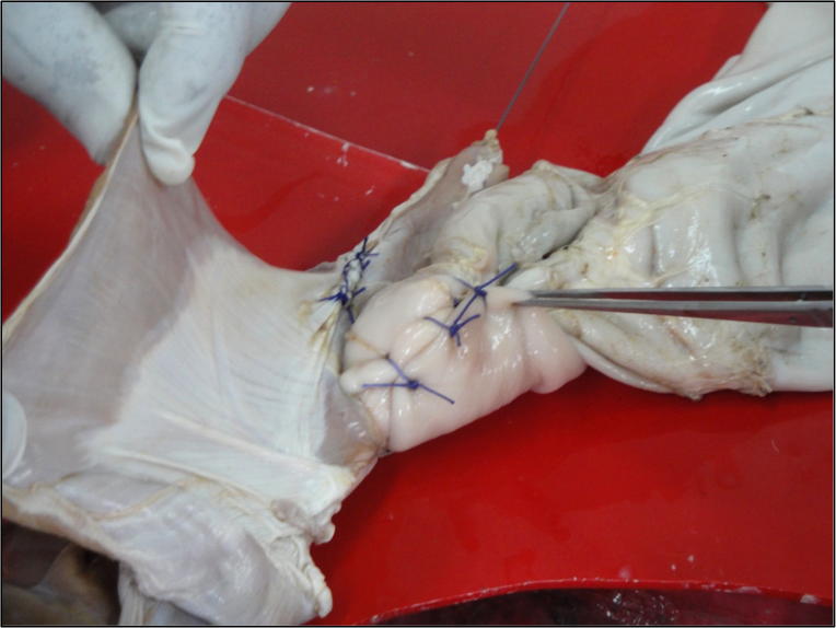
8. Place two “tacking” sutures of 2-0 Ethibond through the “wrap” into the adjacent diaphragm on each side of the wrap (see below).

Figure 2.3: Nissen Fundoplication – Completed 360-Degree Wrap
Video Exercise 2.1
Videos may take a moment to load depending on your connection speed.
Exercise 2.1. Nissen Beginning
Video Exercise 2.2
Videos may take a moment to load depending on your connection speed.
Exercise 2-2. Passing Fundus Posteriorly
Video Exercise 2.3
Videos may take a moment to load depending on your connection speed.