Goals
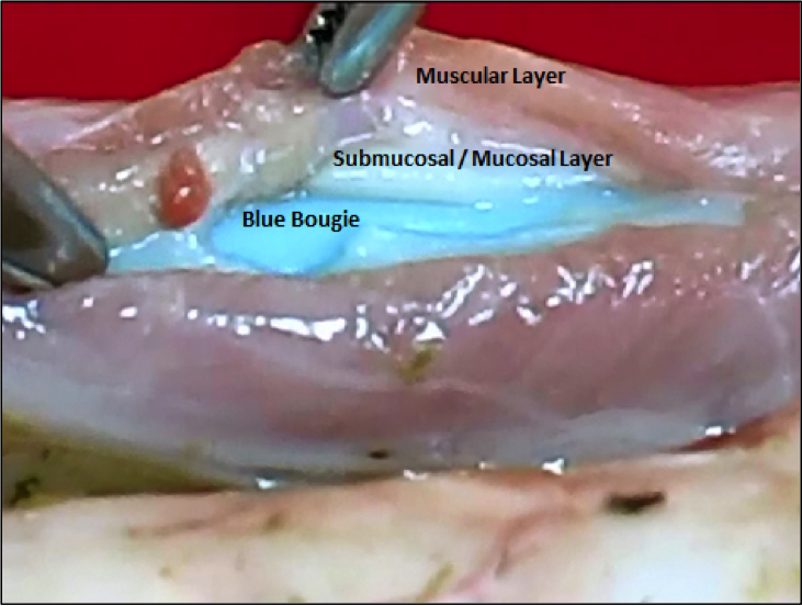
- Observe the phenomenon of the submucosal/mucosal layer laceration extending for a greater distance proximally and distally than the muscular “laceration” and discuss why this is important to know
- Learn the gentle handling of esophageal tissue.
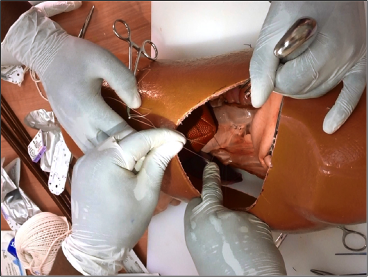
- Experience working through a low postero-lateral thoracotomy incision while repairing a distal esophageal perforation in a patient whose lung is being ventilated on the operative side.
Equipment
- Goat mediastinal tissue block. Fresh from local abattoir. Refrigerate. Consists of heart, lungs, esophagus, trachea, diaphragm, thoracic aorta and esophagogastric junction with attached proximal one third of the stomach.
- Mannequin of human chest (from local department or Men’s store) cut in half along the sagittal plane to create the simulation of a left hemi-thorax. Cut a wedge-shaped hole in lateral side of left chest mannequin to simulate a left postero-lateral thoracotomy incision.
- Two battery operated headlights (from local bike or sports store).
- Maloney tapered bougie—Size 45 French.
- 17-inch x 24-inch plastic cutting board with single screw placed in the center edge of each
end of longest dimension. - Lubricant—tube of K-Y jelly or Lubriderm.
- One pair of Metzenbaum scissors.
- Disposable scalpel with #10 blade.
- 3- 0 vicryl sutures.
- 3-0 silk sutures.
- Long needle holder.
- One pair long surgical pickups.
- Two lung clamps.
- Endotracheal tube.
- Ambu bag for ventilating the lung using the endotracheal tube.
- Small Luer lock syringe to inflate endotracheal tube balloon.
- Two curved hemostats to “tag” sutures placed in esophageal muscle layer for exposure of submucosal layer.
- One lung retractor (eggbeater retractor).
Preparation
- Attach the mediastinal tissue block to the cutting board with #1 Prolene suture passed through the proximal esophagus and trachea. Do not obstruct the esophageal lumen. Tie this suture around the screw at the end of the cutting board. Place a second #1 Prolene suture through the distal diaphragmatic crura and tie around the screw at the distal end of the cutting board.
- Pass a lubricated esophageal bougie the full length of the esophagus.
- Using a scalpel, cut 1-inch longitudinal “laceration” through the esophageal wall down to the bougie.
- Remove the bougie.
- Gently grasp with a forceps the proximal and distal ends of the “laceration” and extend the submucosal/mucosal “laceration” with Metzenbaum scissors 1 cm proximally and distally.
- Insert the endotracheal tube in the trachea, expand the balloon cuff and attach the tube to the Ambu bag.
- Place the chest mannequin over the mediastinal tissue block and put on headlight.
Discussion
Perform Transthoracic Repair of an Acute Esophageal Perforation
1. Identify the site of the esophageal perforation (see below).

Figure 10-1: Esophageal Perforation with Bougie in Esophageal Lumen
2. Gently grasp the muscular layer of the esophagus at the proximal and distal ends of the laceration to assess the extent of the underlying submucosal/mucosal laceration. Extend the muscular layer laceration to expose the ends of the submucosal/mucosal laceration.
3. Close the submucosal/mucosal laceration with interrupted 3-0 absorbable sutures.

Figure 10-2: Tying Sutures to Close the Esophageal Perforation
4. Close the muscular layer laceration with interrupted 3-0 black silk sutures taking care to avoid narrowing the esophageal lumen.
5. Discuss the importance of placing a chest tube in a very dependent position in the chest cavity.
6. Discuss the harvesting of an intercostal muscle pedicle graft for reinforcement of the esophageal tear closure site. Note the importance of suturing the graft to the esophagus in a longitudinal manner. Discuss how wrapping the graft circumferentially around the esophagus will produce a calcified obstructing band around the esophagus due to the fact that the rib periosteal tissue will lay down new calcium.